A new method developed for quantifying complex-formed corals
15 September 2022
Complex branching growth forms occur on a large scale in biology (branching fungi, blood vessel systems, trees), but also in chemistry and physics (branching crystals, electrical discharge patterns). An important problem in characterizing complex branching growth forms is that classical methods are often not very useful for morphometrics. Classical morphometric methods often use so-called `landmarks' (such as the position of the eyes, fins, legs) that are missing in this kind of branching structures.
The coral species Acropora sp. is an important species in modern coral reefs. Particularly in the Indo-Pacific area, a large number of Acropora species occur, which are sometimes difficult to distinguish even by experts. For the monitoring of coral reefs, it is very important to have a method that can analyze the three-dimensional shape of the corals. With such an analysis, different Acropora species can be distinguished and the influence of climate change on the growth process of the coral can also be monitored.
Laser scanner
The 3D images were taken with a laser scanner. The laser scanner is a handheld device that can in principle be used in field studies. The laser scanner takes 3D images of the coral and is a non-destructive method that can also be used to monitor living corals.
The project is a collaboration with Catalina Ramirez-Portilla from the Université Libre. Master student Inge Bieger has done the research as part of her thesis work in the master Computational Science.
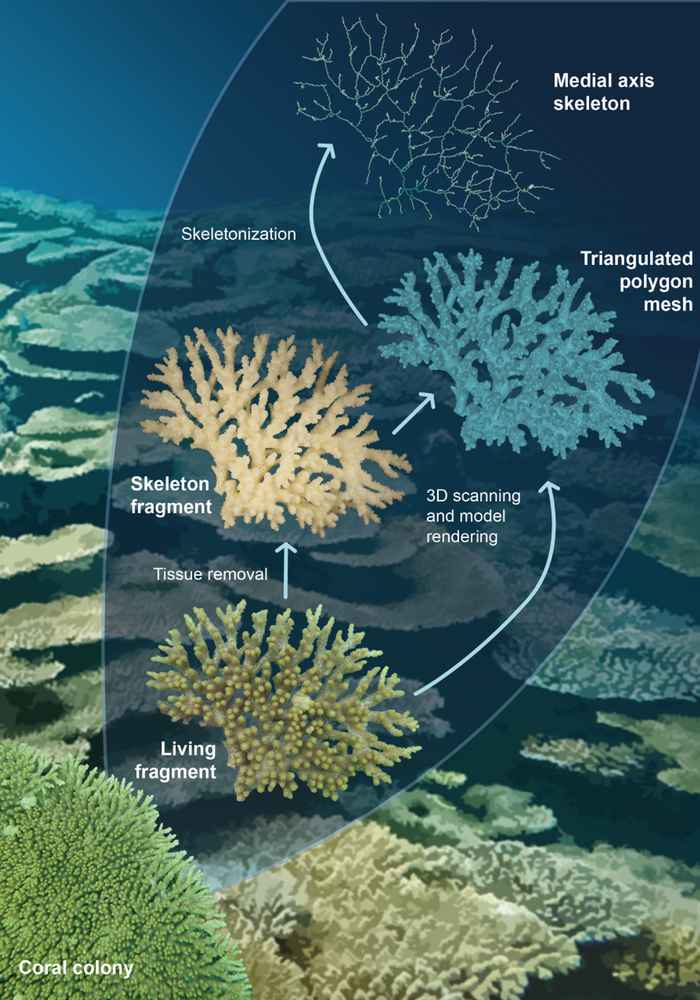
Fig. 1 shows a diagram of this analysis, from the 3D image a branching skeleton is obtained. The branching skeleton is statistically analyzed and can also be used directly in biological interpretations, for example quantifying the degree of contact of the coral with the environment.
An article about this research was accepted for publication in Frontiers in Marine Science.
